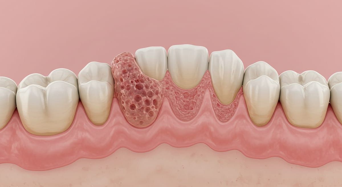
Dental Granuloma: Symptoms, Causes and Treatment
Most often asymptomatic, the dental granuloma can evolve silently for months before causing pain, abscesses or even the loss of the affected tooth. Regularly confused with the dental cyst, the granuloma nevertheless deserves particular attention, because its early management makes it possible to avoid significant complications.
What is a dental granuloma?
A dental granuloma corresponds to a chronic inflammatory reaction that develops at the apex (end) of the root of a tooth. We also speak of an apical lesion.
It is an accumulation of inflammatory cells, sometimes accompanied by a small cavity, which forms in response to a persistent infection inside the root canal. Unlike a malignant tumour, the granuloma is benign, but it never regresses spontaneously.
Over time, the granuloma can evolve into a dental cyst, that is, a cavity surrounded by a membrane and filled with fluid. And it is before this transformation that action should be taken!
Granuloma or dental cyst: what is the difference?
Granuloma and dental cyst present similarities but are not identical.
While the dental granuloma is an inflammatory lesion made up of defensive tissue put in place to contain the infection, the cyst is a closed cavity that gradually enlarges by producing fluid.
In practice, a neglected granuloma can transform into a cyst. Thus, when the dental surgeon detects a granuloma on an X-ray, they must propose a treatment to avoid this evolution. Patients who mention a "cyst" sometimes actually carry an apical granuloma.
What are the symptoms of a dental granuloma?
Most granulomas remain for a long time without apparent manifestation. Several signs can however alert:
- diffuse or persistent pain at the level of a tooth, sometimes accentuated during chewing;
- a feeling of pressure in the jaw;
- abnormal dental mobility;
- localised swelling of the gum;
- the appearance of a fistula (small orifice through which pus flows);
- in some cases, bad breath linked to the presence of bacteria.
These signs are not specific and only an X-ray examination makes it possible to confirm the presence of a granuloma. The image generally shows a dark zone (radiolucent) at the end of the dental root.
What are the origins and risk factors of granulomas?
The formation of a granuloma follows an infectious or traumatic aggression.
Infectious origin
In the vast majority of situations, the dental granuloma results from an untreated cavity or an incomplete root canal treatment. The bacteria proliferate in the root canal and reach the apex. The body reacts by triggering chronic inflammation, intended to contain the infection.
Traumatic or accidental origin
A violent impact, a fracture of the tooth or the jaw can also lead to a necrosis of the dental pulp. In this case, the granuloma develops little by little at the end of the weakened root.
Particular cases of congenital cysts and granulomas
Certain anomalies linked to the development of teeth and maxillae can also favour the appearance of lesions of the granuloma or cyst type. These situations remain rare but justify specific monitoring.
What happens if a granuloma evolves without treatment?
A small granuloma (less than 5 mm in diameter) may go completely unnoticed. But as it grows, several complications can appear.
First of all, the tooth can become painful, move abnormally, or even fall out. The infection can also spread to the surrounding tissues and cause an abscess, swelling of the face or even difficulty eating.
It frequently happens that the evolution leads to a larger dental cyst, with an increased risk of bone loss and weakening of the jaw. This is why it is essential to manage a granuloma as soon as it is diagnosed, even in the absence of marked symptoms.
How to treat a dental granuloma?
The management of the granuloma aims to eliminate the infectious cause and remove the pathological tissues. Two main approaches can be considered, depending on the severity of the lesion and the state of the tooth.
Conservative treatment
When the tooth can be preserved, the dentist carries out a revision of the endodontic treatment (root canal treatment). They clean and disinfect the root canals, before proceeding to their hermetic obturation in order to avoid any reinfection.
In certain cases, a transient antibiotic therapy is prescribed to reduce the inflammation before the resumption of treatment.
Surgical excision
If the granuloma persists despite the root canal treatment, a surgical intervention is required. The excision consists in removing the lesion, as well as the causal tooth when it cannot be preserved.
This operation, called "apicectomy" (or apical resection), is limited to the end of the root which is sectioned. It is performed under local anaesthesia and remains minimally invasive. Bone healing is generally quick.
In every case, the treatment of the granuloma must be accompanied by management of the initial cause, failing which the lesion will eventually recur.
In certain situations of surgical failure, or when the tooth is too mobile or too damaged, a tooth extraction will be necessary.